A fascinating study from researchers at the Centre for Genomic Regulation (CRG) in Barcelona has uncovered an intriguing survival mechanism in cancer cells when they face mechanical stress. This discovery could shift the way we approach cancer treatment.
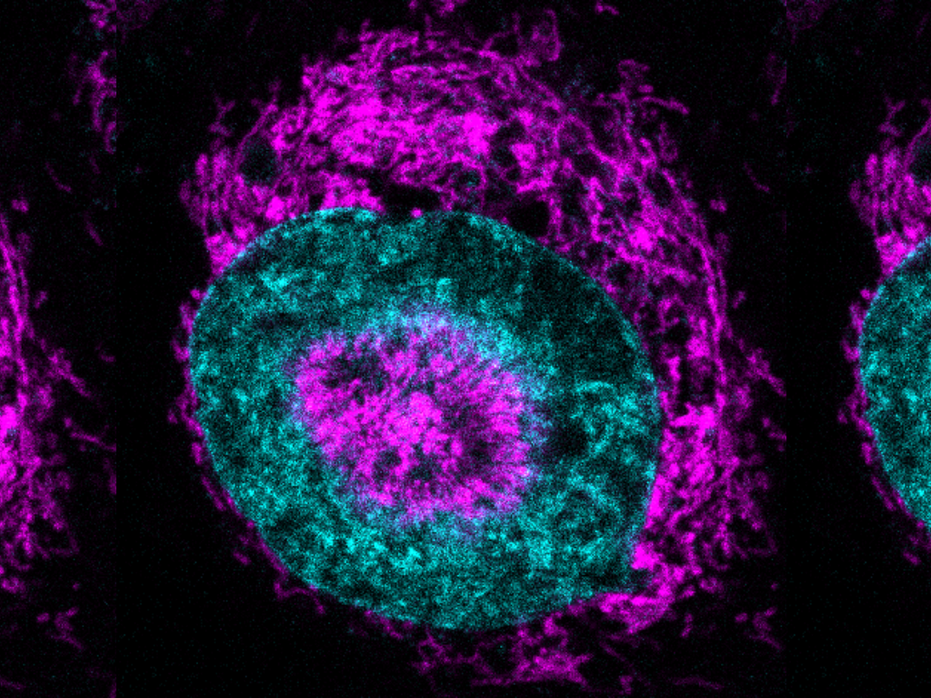
When cancer cells are compressed, their mitochondria—the energy powerhouses—rapidly mobilize towards the nucleus, forming clusters known as nucleus-associated mitochondria (NAMs). This movement causes a swift release of adenosine triphosphate (ATP), an essential energy molecule, directly into the nucleus, providing a burst of energy crucial for DNA repair. In just three seconds, this ATP level spikes by about 60%, playing a critical role in repairing DNA, which is under stress due to compression. Without this energy boost, cells struggle to divide properly.
This entire process hinges on the cell’s internal scaffolding, made up of actin filaments, and involves the endoplasmic reticulum, which helps trap mitochondria near the nucleus. Disrupting this support system with latrunculin A, a compound that breaks down actin filaments, can prevent NAM formation and halt the ATP surge.
Analyses of breast tumor samples from 17 patients showed a marked increase in NAM presence at the tumor’s invasive edges compared to its dense core. This suggests that manipulating the cell’s internal framework could potentially limit a tumor’s invasiveness while sparing healthy tissue.
The researchers used advanced microscopy to compress cells to just three microns wide, observing this remarkable phenomenon in 84% of compressed HeLa cells—an immortal line of human cervical cancer cells known for their use in scientific research.
This groundbreaking work not only advances our understanding of how cancer cells endure mechanical stresses but also opens the door to potential new therapeutic targets. By focusing on this energy response mechanism, future treatments might more effectively curb cancer’s invasive spread.